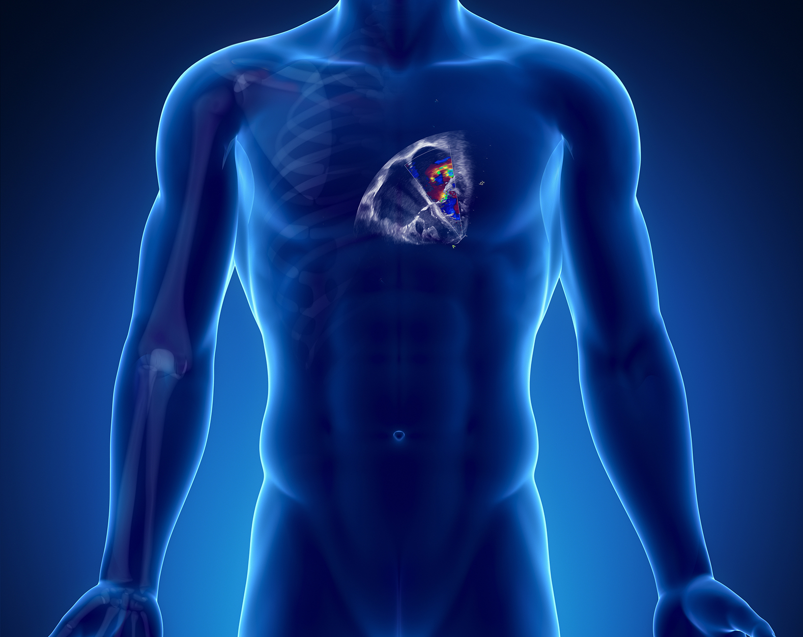
Echocardiography
Keeping cardiology and vascular ultrasound exams well organised and easily accessible is essential in today’s busy echo departments. With reporting and archiving capabilities, ViewPoint 6™ with EchoPAC™ Suite can help streamline and simplify your ultrasound workflow.

Streamline ultrasound reporting and image management for Echocardiography
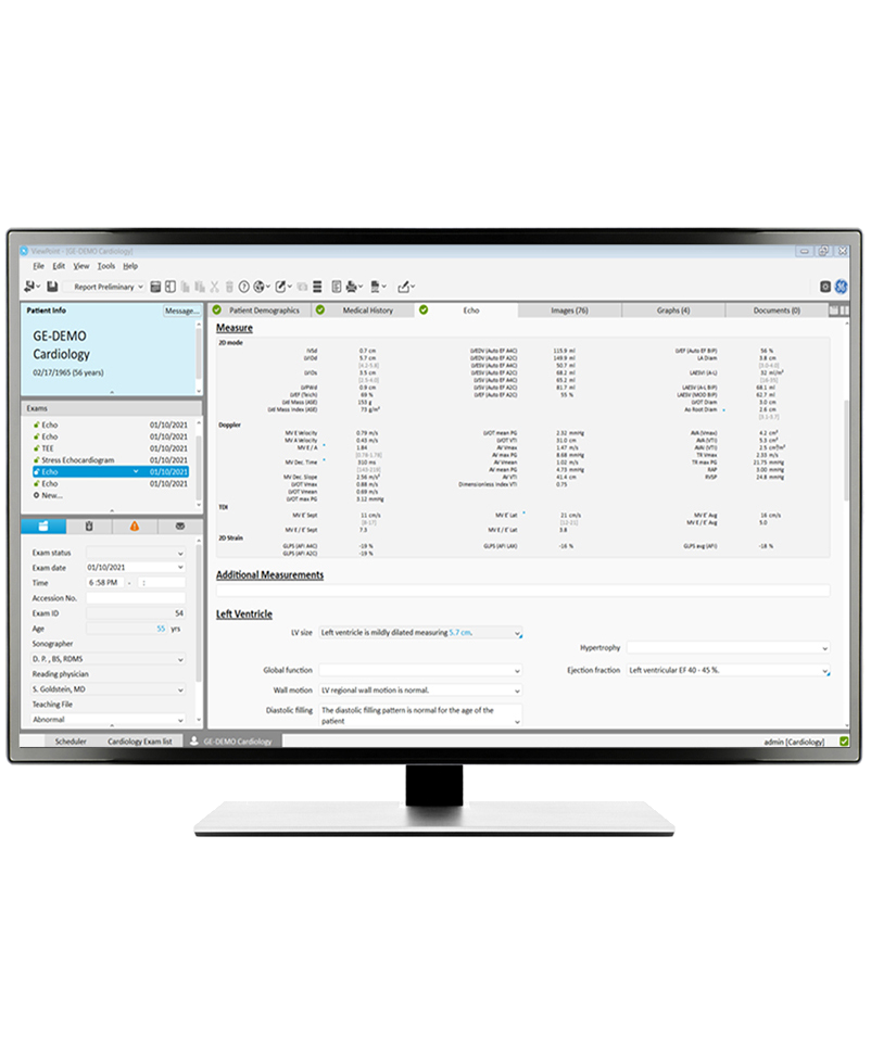
ViewPoint 6 has been designed to simplify image management, reporting, workflows and data gathering in hospitals and private practices. Patient data, detailed structured exam findings and images are available together in a single view. Existing data and imaging from legacy systems such as Image Vault™ can be migrated to ensure all data remains available in a single system.
ViewPoint 6 for Echocardiography content
Choosing ViewPoint 6 for all your echocardiography content means being able to store:
Transthoracic echocardiography.
Transesophageal echocardiography.
Stress echocardiography.
Paediatric transthoracic echocardiography.
Paediatric transesophageal echocardiography.
Professional Reporting
With reporting modules developed specifically for echocardiography you can expect features such as:
Organisation-wide, fast, comprehensive documentation of ultrasound exams.
Automatic transfer of measurements from the ultrasound modality or EchoPAC Suite directly into the report.
Quick Report: timesaving documentation of standard findings.
Tailor reporting forms to local requirements.
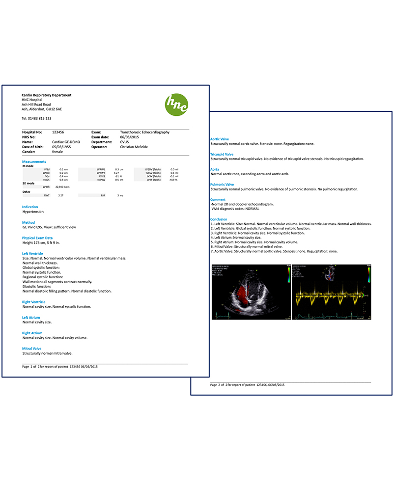
Include images, charts, and drawings on the report.
Export report to third-party systems in PDF or DICOM format.
Switch between the display of the exam form and the report preview with one click.
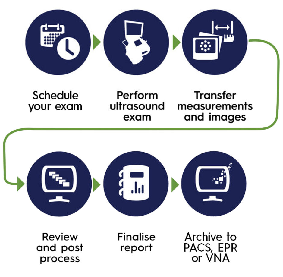
Simple Workflows
Integrate ViewPoint 6 with existing hospital systems such as PAS, RIS, EPR, PACS, VNA to streamline workflows.
Automatic image and measurement transfer to third-party systems.
Statistical analysis of the database with user-defined queries.
Import of documents into an exam.
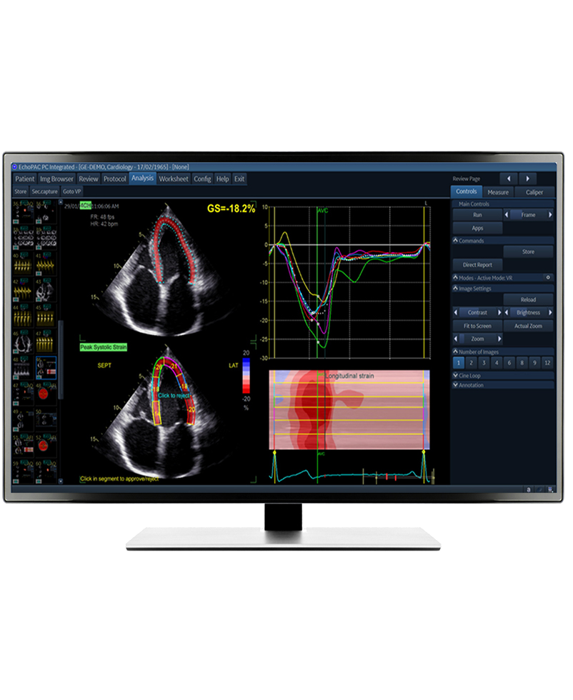
Comprehensive Image Management
Installing ViewPoint 6 with the EchoPAC Suite enables you to review images, DICOM multiframe and Vivid raw data from a study in one place.
Other features include:
Measurements and Analysis.
4D and Multi-dimensional Imaging.
Stress Echo.
Auto Ejection Fraction (EF).
Automated Function Imaging (AFI).
Tissue Synchronization Imaging (TSI).
2D Strain.
Myocardial work.
Advanced Qscan Imaging.
4D Auto Aortic Valve Quantification.
4D Auto Mitral Valve Quantification.
4D Auto Tricuspid Valve Quantification.
Intima Media Thickness (IMT).
4D Auto Right Ventricle Quantification.
4D Auto Left Atrium Quantification.
Measurements available in report immediately.
Image transfer of single images or video sequences from ultrasound systems and other DICOM modalities.
Integrated EchoPAC Suite for post-processing volumes from GE Vivid modalities.
Export of anonymised images with comments for teaching and presentations.
Select images to be included in the printed report.
Customisable display of images and graphs.
Auto-play of image sequences.
Feature to compare images between exams.
Remove need for printing images on thermal paper.

Information at your fingertips
The integrated analytics module in ViewPoint 6 can help put all that data at your fingertips to look for trends and answers to clinical and administrative questions.
Create queries easily for local and national returns, workflow enhancements, trending and research.
Run queries and filter on any field in the database.
Analyse measurements.
Query free-text and fixed answers.
Export to other applications such as Microsoft™ Excel™ with a single click.